
Mechanism of Interneural Edema in Carpal and Cubital Tunnel
Filed under Diagnoses
Mechanism of Interneural Edema
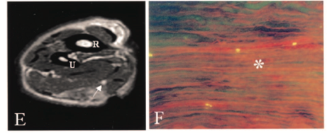
Over the last few weeks I have been learning about ultrasonic imaging and carpal tunnel syndrome. When reviewing carpal tunnel syndrome, I learned that intraneural edema is a common sign of compression injuries such as carpal tunnel and cubital tunnel. There are numerous causes of carpal tunnel syndrome, and every scenario ends with the reduction of available space within the carpal tunnel and the inevitable compression on the median nerve (carpal tunnel edema). What I did not know was that chronic compression on the nerve can disrupt and open the blood nerve barrier around the perineurial layer. This allows for blood to flow freely into the nerve causing swelling or interneural edema. Since the nervous system lacks lymphatic drainage in the endoneural space, swelling inevitably increases pressure and disrupts the flow of blood to the nerve resulting in a metabolic conduction block (Cooper, 2014). One animal study found that an increase in pressure as little as 30 grams of force (about the weight of an average lightbulb) over the course of 1 hour was enough to disrupt the blood nerve barrier around the median nerve and cause diffusion (Kobayashi et al., 2005).
Normal Nerve No Diffusion
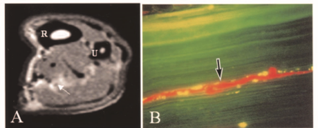
Nerve 30 Grams of Force with Diffusion
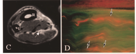
Nerve 90 Grams of Force Severe Diffusion
Chronic compression and decreased blood flow lead to impairment in nerve conduction. One source states that functional deficits are seen sequentially in the following order: motor, proprioception, touch, temperature, pain, and then sympathetic function (Cooper, 2014). Therapeutic activities such as nerve gliding exercises are hypothesized to increase nerve mobility and release the nerve from the sight of compression. Additionally, surgical decompression can help to alleviate symptoms, but the timeline for neural repair is largely based on the severity of nerve damage that has occurred. As neural edema subsides and blood flow to the nerve improves, the nerve begins to repair itself as long as the endoneurial tubes are intact. Patients are expected to regain sensation in the reverse order that they were initially lost (pain, temperature, proprioception).
Cooper, C. (20014). Fundamentals of hand therapy: Clinical reasoning and treatment guidelines for common diagnoses of the upper extremity [Second Edition]. Elsevier Mosby
Kobayashi, S., Meir, A., Baba, H., Uchida, K., and Hayakawa, K. (2005). Imaging of intraneural edema by using gadolinium-enhanced MR imaging: Experimental compression injury
2 Comments
Leave a Comment
More To Read

7 Tips for your Osteo Arthritis Patients!
7 Tips for your OA Patients! Managing Osteoarthritis in the Hand Our hands are one of the most intricate structures in the human body. They are composed of a network of tendons, ligaments, and nerves that make it possible to perform daily tasks such as unlocking a door, peeling an egg, or sending an email…

Hand Therapy: How to Treat the Client with a New Distal Radius Fracture
A short blog post on the basics of treating a Distal Radius Fracture.

Do you know the difference between an Electromyography (EMG) and a Nerve Conduction Velocity (NCV) Study?
Do you know the difference between EMG and NCV (an Electromyography and a Nerve Conduction Velocity Study? The term nerve test is usually a broad term that typically indicates both an Electromyography (EMG) and a Nerve Conduction Velocity (NCV) study (EMG vs NCV). An EMG looks at the electrical signals your muscle makes when at…

Stretching Alone Can Change P1 Bone Shape in Patients with Camptodactyly
Woo Hong, S. Kim, J., Sang Kwon, O., Ho Lee, M., Sik Gong, H., Hyun Baek, G., (2019). Radiographic Remodeling of the Proximal Phalangeal Head Using a Stretching Exercise in Patients With Camptodactyly. J Hand Surg Am, 1.e1-1.e10 The Skinny – Camptodactyly is a congenital, nontraumatic flexion contracture of the PIP in fingers other than…

Sign-up to Get Updates Straight to Your Inbox!
Sign up with us and we will send you regular blog posts on everything hand therapy, notices every time we upload new videos and tutorials, along with handout, protocols, and other useful information.
The inter neural edema is fascinating.
Yes is is very interesting! I agree